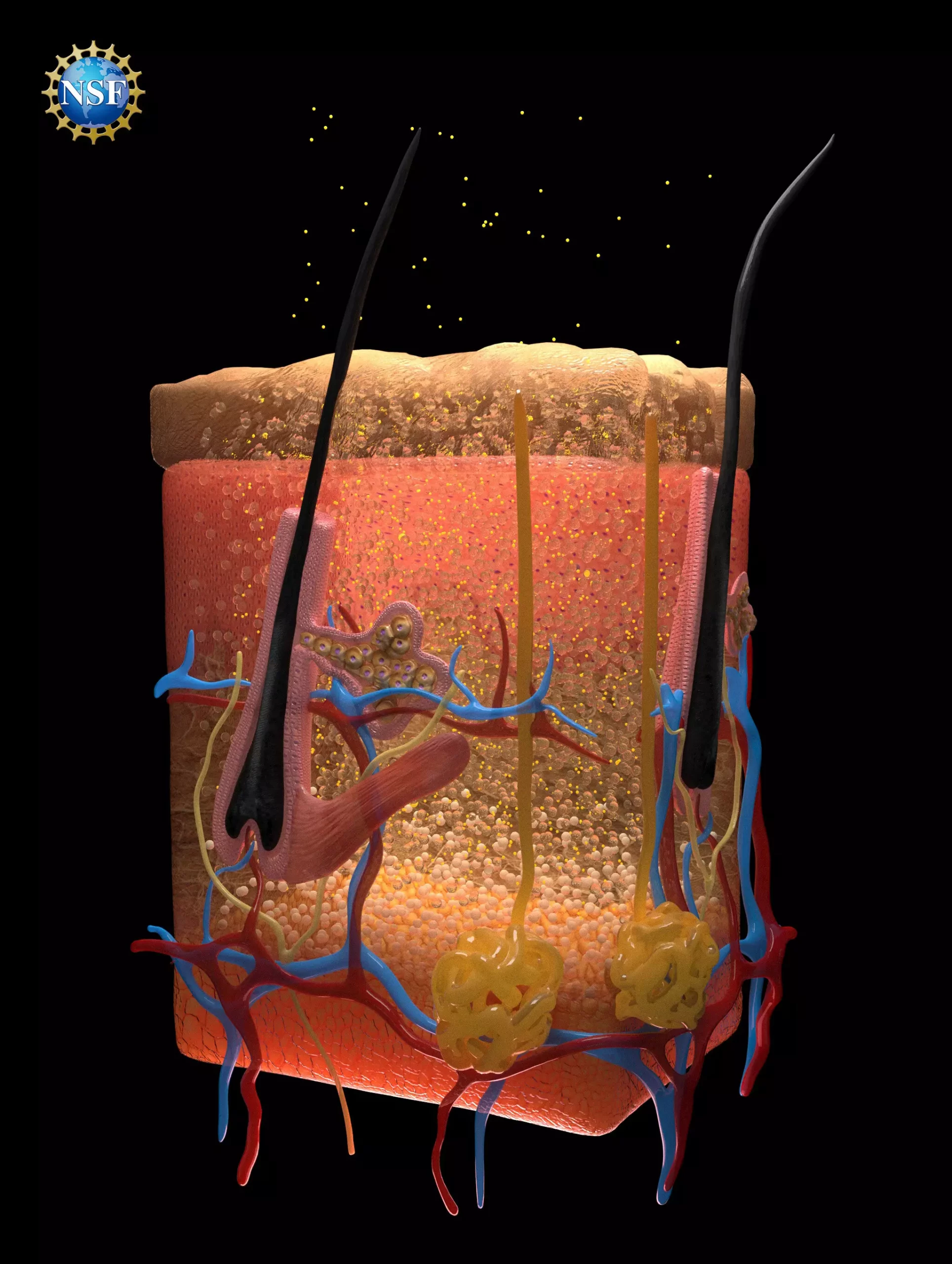
In a remarkable advancement in medical diagnostics, researchers at Stanford University have unveiled a novel method that allows for unprecedented visibility of internal organs by rendering overlying tissues transparent. This innovative process hinges on a seemingly simple application of a food-safe dye, yet its implications for the medical field are vast and multifaceted. The research, which was published in the prestigious journal Science, presents a promising approach for improved visibility in various medical scenarios, such as identifying cancers, monitoring digestive disorders, and facilitating blood draws.
At the crux of this technique is the understanding of how light interacts with biological tissues, particularly focusing on light scattering and the concept of refraction. Biological tissues are composed of fats, fluids, proteins, and other materials, each contributing to different refractive indices. This variance in refractive indices leads to light scattering, which, in turn, gives biological tissues their opaque appearance. The researchers recognized that to achieve transparency, it was essential to match these refractive indices, allowing light to pass through without disruption.
Through meticulous experimentation, the team discovered that certain dyes, specifically tartrazine (common food dye), could not only absorb light but also create uniformity in the refractive indices of surrounding biological materials. By dissolving tartrazine in water and applying it to biological tissues, they demonstrated a significant increase in transparency as the dye effectively altered the way light interacted with the tissue.
Applications in Medical Diagnostics
The potential applications of this technique are revolutionary. Imagine a healthcare setting where veins and arteries are made more visible for blood draws, or where laser treatments for cancer become more effective due to improved light penetration. The implications extend to a myriad of diagnostic and therapeutic processes. For instance, utilizing this technique in combination with existing laser therapies could enhance the effectiveness of treatments targeting tumors located deeper within the body, thereby improving patient outcomes.
In their experiments, researchers first validated their predictions using thin slices of chicken breast, where increased concentrations of tartrazine produced a transparent effect. Subsequently, the solution was applied to live animal subjects, revealing intricate blood vessels and internal organ movements. The reversibility of the process is equally significant; tissues return to their original opacity promptly after rinsing, suggesting that the technique is safe and non-invasive.
The researchers embarked on this project with a fundamental question about how microwave radiation interacts with biological tissues. In delving into this inquiry, they unearthed long-standing principles from optics, such as the Kramers-Kronig relations, which provide crucial mathematical frameworks for understanding light behavior. This foundational knowledge was instrumental in devising a method to tailor dyes that enhance optical clarity in biological tissues, ultimately leading to the formulation of the transparency technique.
Graduate researcher Nick Rommelfanger and postdoctoral researcher Zihao Ou spearheaded the initiative, collaborating with a dynamic team of 21 individuals—including students and advisors—dedicated to exploring the optical properties of various dyes. The research took advantage of both traditional and cutting-edge analytical equipment, including an ellipsometer, a tool typically utilized in semiconductor manufacturing rather than biological imaging. This unconventional application highlights the interdisciplinary nature of the research, showcasing how established tools from one field can find transformative uses in another.
Looking ahead, the researchers aspire to broaden the scope of their findings by exploring the potential for injecting dyes to access even deeper layers of biological tissues. The prospect of developing customized dyes tailored to match the optical requirements of specific tissues could pave the way for unprecedented clarity in medical imaging.
The team’s efforts underscore a fundamental shift in how we approach biomedical research. By blending cutting-edge optical science with practical applications in medicine, this work fosters optimism for future innovations that could redefine diagnostic processes. As the field of medical optics progresses, we may soon witness a new era of non-invasive imaging techniques, fostering a better understanding of human anatomy and enhancing the ability to diagnose and treat medical conditions at earlier stages.
The development of the transparency technique marks a significant milestone in medical research. The ability to visualize internal organs without invasive procedures holds the potential to revolutionize diagnostics and treatment methodologies across various medical domains. By merging fundamental scientific principles with practical medical applications, this research opens new avenues for understanding human biology and treating diseases more effectively.


Leave a Reply