
In the realm of cellular biology, understanding the intricate structures and functions within cells is fundamental to numerous scientific advancements. Historically, conventional microscopes have been restricted in their ability to provide detailed images of cellular components due to resolution limits. With resolutions typically starting from around 200 nanometers, many cellular structures, particularly those within human cells, have eluded scientists’ scrutiny. For example, the delicate filamentous scaffold within cells measures a mere seven nanometers in width, and the synaptic cleft between nerve cells spans just 10 to 50 nanometers. These minute dimensions pose significant challenges, as standard microscopes can only deliver fragmented images, hampering our ability to fully comprehend the cellular landscape.
However, a groundbreaking development from researchers at the Universities of Göttingen and Oxford, in partnership with the University Medical Center Göttingen (UMG), is set to transform this field. They have introduced an innovative microscope that achieves a remarkable resolution better than five nanometers—an astounding feat represented by the metaphor of dividing a single hair into 10,000 strands. Such enhanced imaging capabilities catalyze a surge in our understanding of cellular structures, providing clearer, more comprehensive insights into the microcosm of life that exists at the nanoscale.
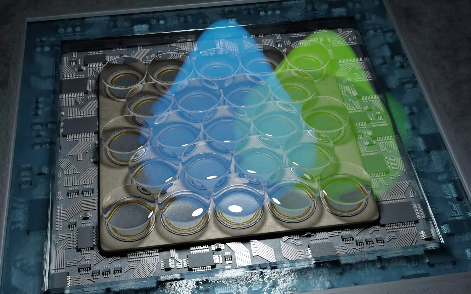
This novel approach leverages a specific type of microscopy called fluorescence microscopy, which operates through a technique known as “single-molecule localization microscopy.” This methodology involves the precise activation and deactivation of individual fluorescent molecules within a sample, allowing scientists to pinpoint their exact positions with unparalleled accuracy. By modeling the entire structure based on these localized positions, researchers can now uncover intricate details previously hidden from view.
The research team, led by the esteemed Professor Jörg Enderlein from the University of Göttingen’s Faculty of Physics, has achieved remarkable advancements in resolution capability, effectively doubling previous limits. Utilizing a cutting-edge, highly sensitive detection system along with sophisticated data analysis techniques, this newly developed microscope can reveal minuscule details, such as the specific organization of proteins at synapses between nerve cells. Such insights are not merely academic; they hold the potential for profound implications in understanding neurobiology and cell signaling.
As Professor Enderlein articulates, the new technology represents a pivotal advancement in high-resolution microscopy, offering unparalleled resolution while simultaneously being cost-effective and user-friendly. This is of paramount importance, as accessibility often governs the dissemination of scientific methods across various disciplines and research settings.
In an encouraging move towards broadening access to this groundbreaking technology, the research team also developed an open-source software package designed for the processing of data acquired through their new microscopy method. This initiative is a significant step toward democratizing access to advanced imaging techniques, facilitating utilization by a diverse array of specialists, researchers, and educational institutions. The implications of such accessibility are immense; it fosters collaboration, encourages interdisciplinary studies, and accelerates scientific discovery across multiple fields within the life sciences.
Moreover, the potential applications of this high-resolution microscopy extend beyond fundamental research. It can significantly enhance our understanding of disease mechanisms, provide insights into cellular dysfunction, and contribute to the design of targeted therapeutics. As this technology continues to evolve, its ripple effects will likely be felt across various domains within biology and medicine.
As we stand at the precipice of a new era in microscopy, the impact of this research holds enormous promise. The ability to visualize cellular structures with such precision enables not only a deeper understanding of cell biology but also strengthens the foundation for future innovations in medicine and biotechnology. As researchers continue to push the boundaries of what is possible in imaging technology, we may soon find ourselves uncovering even deeper layers of the cellular world, all thanks to the transformative power of nanoscopic microscopy. The journey into the infinitesimal realms of life is just beginning, and the discoveries yet to come are bound to be nothing short of extraordinary.
Leave a Reply