Recent advancements in neuroscience have brought the intricate workings of neural circuits into sharper focus. One of the pivotal tools in this exploration is genetically encoded voltage indicators (GEVIs), which allow researchers to visualize electrical activity in neurons. Understanding how neurons communicate through electrical signals is vital for deciphering the brain’s complex coding mechanisms. However, as scientists delve deeper, they face the critical task of optimizing their imaging techniques, particularly the perennial debate between one-photon (1P) and two-photon (2P) voltage imaging methodologies.
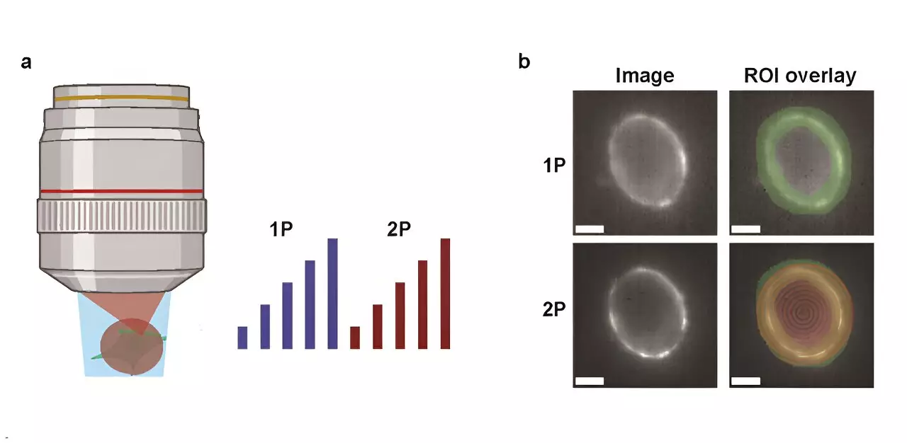
A recent study conducted by a team at Harvard University has provided significant insights into the comparative efficacy of 1P and 2P voltage imaging techniques. Published in the journal Neurophotonics, the researchers meticulously compared these two imaging methods, analyzing their respective optical properties and biophysical limitations. This comprehensive exploration centered on key attributes like brightness and voltage sensitivity of GEVIs while examining how fluorescence diminishes with depth within the murine brain—an essential consideration for in vivo imaging.
The study introduced a predictive model to estimate the number of detectable cells, taking into account important factors such as the properties of the reporters, imaging settings, and the necessary signal-to-noise ratio (SNR). This modeling is a significant step forward in quantifying the capabilities of each imaging strategy.
One of the striking revelations from the research is the substantial discrepancy in power requirements for 1P and 2P excitation. Specifically, it was found that 2P imaging necessitates approximately 10,000 times more illumination power per cell than its 1P counterpart to achieve equivalent photon count rates. This enormous lighting requirement can lead to various complications, such as increased risk of photodamage to tissue and elevated shot noise levels, which ultimately hampers the quality of imaging results.
For example, the application of the JEDI-2P indicator within the mouse cortex revealed that with a targeted SNR of 10, a measurement bandwidth of 1 kHz, and a laser power constraint of 200 mW, researchers could only effectively monitor around 12 neurons when looking beyond a depth of 300 micrometers. Such limitations underline the inherent difficulties of conducting 2P voltage imaging in a living organism.
The findings of this research indicate that while 2P imaging might offer certain advantages, its current manifestations come with significant obstacles. The study emphasizes that enhancing SNR capacity for a higher number of neurons at greater depths will require either groundbreaking advancements in 2P GEVIs or entirely novel imaging methodologies.
Understanding the trade-offs between 1P and 2P imaging techniques will continue to ignite discussions and innovations within the scientific community. As researchers endeavor to push the boundaries of voltage imaging, the lessons derived from this study will serve as a critical guide for future technological developments. The quest to unlock the secrets of the brain’s neural circuits depends not only on current methodologies but also on a commitment to advancing these essential tools in neuroscience.


Leave a Reply