
In the realm of scientific exploration, breakthroughs in imaging techniques can pave the way for unprecedented advancements across various fields. A remarkable innovation has unfolded at Trinity College Dublin, where a group of international scientists has charted a new course in microscopy. By devising an advanced imaging method that significantly minimizes both the time and radiation exposure required for high-quality images, this research stands to impact disciplines ranging from materials science to biomedical research. It highlights a transformative moment that breaks down longstanding barriers in microscopy and sets a new standard for studying delicate biological samples.
Understanding the Conventional Process
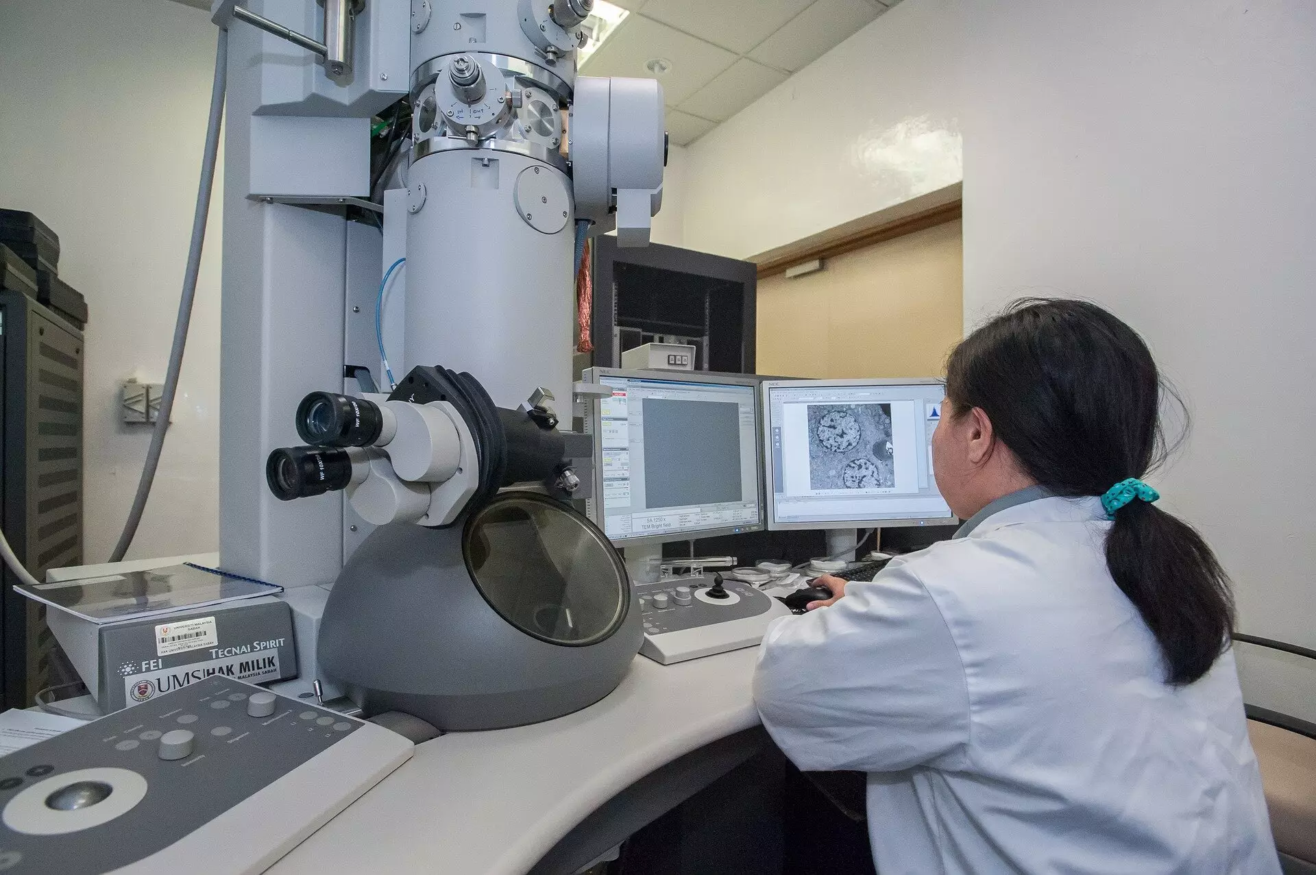
To fully appreciate the significance of the researchers’ findings, it is crucial to understand the traditional paradigm of scanning transmission electron microscopy (STEM). Traditionally, the imaging process involves directing a finely focused electron beam across a specimen, gathering data point by point. In this conventional method, the beam remains fixed on each pixel for a predetermined duration, a practice that parallels the operation of old-fashioned cameras using photographic film. While this straightforward approach has served the scientific community for decades, it comes with significant drawbacks. Specifically, prolonged exposure to the electron beam can inflict damage on sensitive materials, such as biological tissues, which require meticulous care during examination.
As scientists continuously bombard specimens with electrons, the risk of transformation or destruction looms large. This trade-off between obtaining detailed images and safeguarding specimens will resonate with many in the scientific community; the results can be frustrating, producing images that are either unusable or misleading in critical research.
Redefining Imaging with an Event-Based System
The groundbreaking innovation from Trinity College challenges the status quo by radically rethinking how imaging is approached at a fundamental level. Moving away from the standard fixed dwell time per pixel, the researchers introduced an event-based detection system. In this model, the efficiency of imaging is measured by the time taken to gather a set number of scattered electron events rather than the total count gathered over a fixed duration. This paradigm shift allows scientists to gather more useful information from the very first electron detected at any given point, suggesting diminishing returns from subsequent electron impacts.
By harnessing the new theoretical framework, the research team discovered a way to optimize imaging efficiency. The implications are profound: they can now ‘shut off’ the electron beam right at the moment peak efficiency is achieved, significantly decreasing the requisite radiation dose needed to produce maximal image quality. This elegant solution addresses a pressing challenge in the field of microscopy, serving both the need for high-resolution visuals and the imperative to protect sensitive biological materials.
Technical Implementation and Real-World Impact
The application of this novel theory forms the backbone of the patented Tempo STEM technology, developed in collaboration with IDES Ltd. At the heart of this innovation lies a sophisticated “beam blanker,” which allows for the rapid on-off modulation of the electron beam in real-time. This means that instead of bombarding the specimen with excessive electrons, researchers can now fine-tune their approach, shining light on the importance of methodical analysis in microscopy.
Dr. Lewys Jones, an esteemed figure in the field and a lead researcher on the project, articulates the significance of this advancement: “Combining two already state-of-the-art technologies in such an exciting way delivers a real leap in the microscope’s capabilities.” The ability to control the electron beam with nanosecond precision in response to real-time events marks a notable advancement that could redefine how microscopy is practiced. As Dr. Jon Peters pointed out, while electrons are often perceived as harmless in comparison to other forms of radiation, their high velocity can cause substantial damage at a microscopic level, making this innovation not only exciting but crucial for accurate scientific analysis.
The Future of Microscopy
In an age where precision and accuracy are paramount to scientific integrity, the developments emerging from Trinity College Dublin are not merely incremental improvements; they represent a seismic shift that could influence future research methodologies. This pioneering work promises not only to enhance the quality of microscopic imaging but also to protect the integrity of samples that are critical for groundbreaking studies in biology, medicine, and materials science. The ramifications of this research will no doubt resonate widely, encouraging a new wave of inquiry that prioritizes both quality and safety in scientific exploration.
Leave a Reply