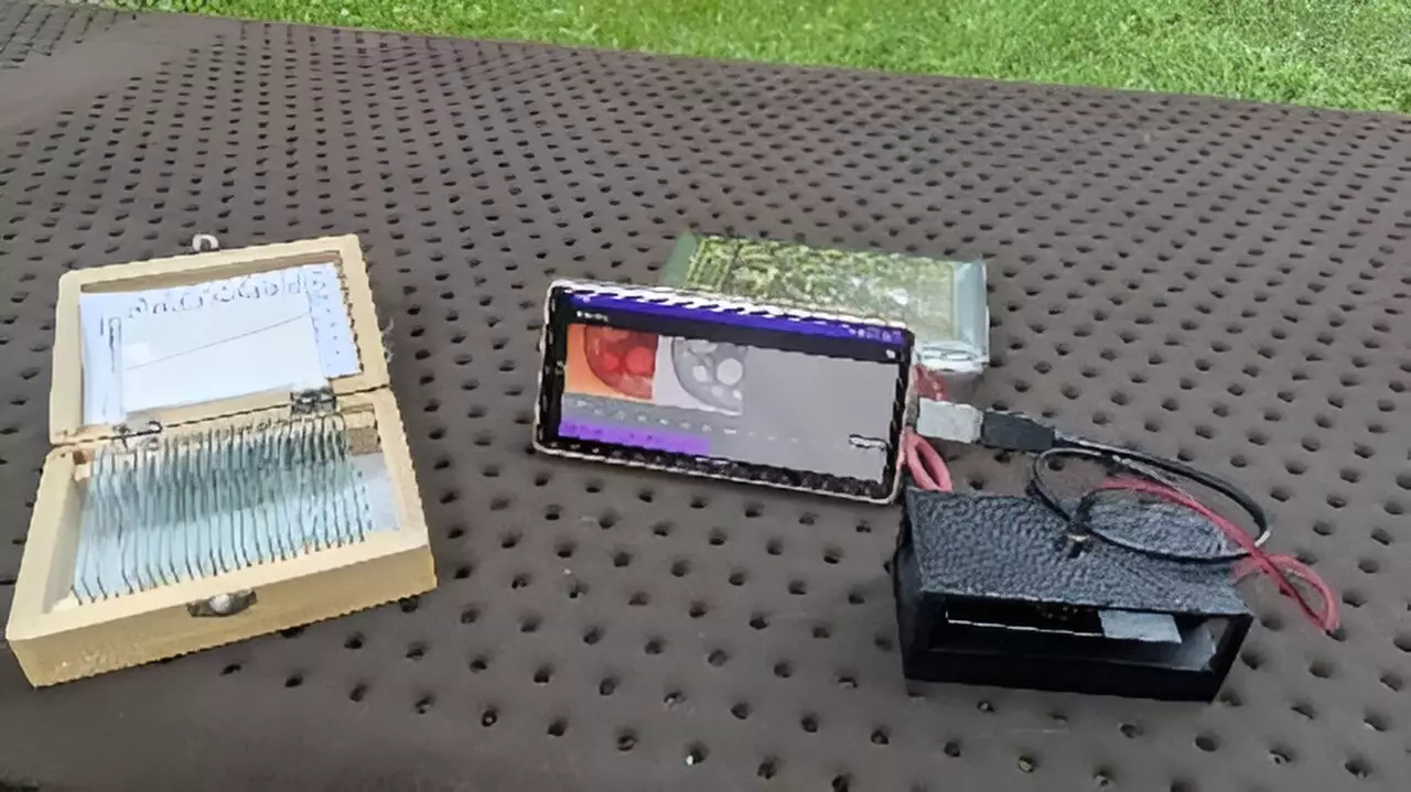
In recent years, advancements in microscopy have ushered in a new era of research and diagnostics. One particularly exciting development is the creation of a smartphone-based digital holographic microscope. This innovative technology holds the potential to democratize access to advanced imaging techniques, bringing 3D measurement capabilities within reach of diverse applications, ranging from education to critical medical diagnostics. This article delves into the significance, functionality, and future implications of this remarkable tool.
The integration of smartphones in scientific research signifies a paradigm shift from traditional microscopy approaches, which are often cumbersome and costly. Conventional digital holographic microscopes rely on intricate optical configurations and necessitate substantial computational resources, typically tied to personal computers. This reliance restricts their mobility and accessibility, particularly in settings where resources are constrained, such as schools in underprivileged areas or healthcare facilities in developing nations.
The new smartphone-based microscope changes this narrative. As Yuki Nagahama, lead researcher from the Tokyo University of Agriculture and Technology, highlights, “Our digital holographic microscope uses a simple optical system created with a 3D printer and a calculation system based on a smartphone,” paving the way for a more affordable and versatile tool. This innovative system not only enhances portability but also ensures that advanced 3D measurements can be conducted in real-time, transforming how microscopy can be utilized in various fields.
The operational principle behind digital holographic microscopy lies in its ability to capture and reconstruct holograms, facilitating the extraction of sophisticated 3D data concerning specimens. By employing an interference pattern generated by a reference light beam juxtaposed with light scattered from the specimen, this technology can unveil intricate details about the sample’s internal structure as well as its surface characteristics.
Previous attempts at developing smartphone-based holographic microscopes encountered hurdles, notably the limitations in processing capacity and memory that hindered real-time image reconstruction. However, the innovative approach of utilizing band-limited double-step Fresnel diffraction has proven to be a game changer. This technique streamlines data processing by significantly reducing the volume of information that must be computed, thereby enabling faster and more efficient hologram reconstructions directly on smartphones.
A critical advantage of this new digital holographic microscope is its lightweight design emerging from 3D printing technology. This not only minimizes manufacturing costs but also enhances the practicality of the device across various environments. The exploration of educational realms, for instance, allows students to engage with live biological specimens both within the confines of a classroom and in home environments, enriching their learning experience through hands-on observation.
Furthermore, the medical applications of this technology are monumental. Disease diagnosis, such as sickle cell anemia in developing regions, can benefit from the ease of use and affordability of this smartphone microscope. As Nagahama pointed out, the microscope’s potential in medical settings could help bridge the gap in healthcare accessibility, equipping healthcare providers with advanced diagnostic capabilities without the need for costly laboratory infrastructure.
The operational capabilities of this smartphone microscope extend beyond conventional imaging. With the seamless integration of software applications, users can utilize gestures to manipulate hologram images on their phone screens, enhancing interaction and observational versatility. Initial evaluations demonstrated promising results, allowing for the retrieval of known patterns and the imaging of organic structures, showcasing its practical effectiveness.
Moreover, ongoing research aims to further refine the imaging quality of this innovative microscope by leveraging deep learning techniques. This intersection of artificial intelligence and microscopy could address issues such as the appearance of unwanted artifacts in reconstructed images, ultimately enhancing diagnostic accuracy and the clarity of visualizations.
The development of a smartphone-based digital holographic microscope presents an extraordinary leap toward making high-tech microscopy accessible and viable for a wide array of sector applications. From education to healthcare, this emerging technology augments traditional methods with significant advantages in portability, affordability, and user interaction. The integration of deep learning into this microscope may further bolster its effectiveness, blurring the lines between sophisticated laboratory instrumentation and daily usability. As researchers continue to push the boundaries of what is possible, the future of microscopy looks brighter than ever, paving the way for unprecedented opportunities across scientific disciplines.


Leave a Reply